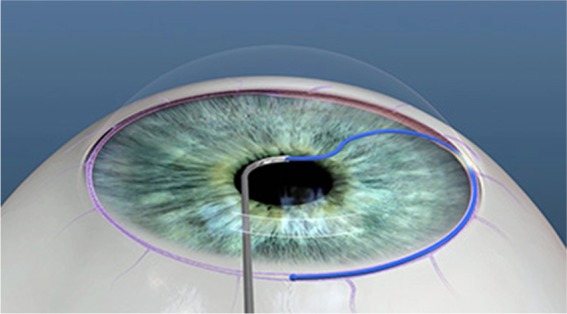
Figure 2.

Cannula tip of the device is used to gain access into Schlemm’s canal.
Notes: The figure depicts the microcatheter advancing into and around Schlemm’s canal. The process can be repeated in an opposite direction for the remaining 180° of the trabecular meshwork. The microcatheter is withdrawn to unroof Schlemm’s canal providing a 360° trabeculotomy. Reproduced with permission from Sight Sciences, Inc., http://sightsciences.com.23
