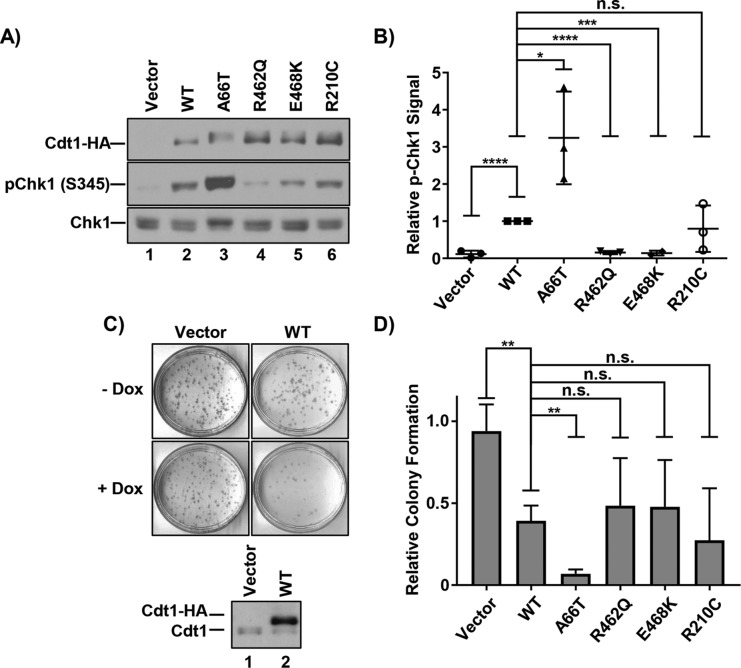
FIGURE 2:
DNA damage and cell proliferation defects from Cdt1 variant overproduction. (A) Immunoblots of HA-tagged Cdt1 (anti-HA antibody), pChk1 (S345), and total Chk1 in U2OS cells grown in 1 µg/ml dox for 48 h. (B) Graph of pChk1 (S345) induction normalized to WT Cdt1. Bars represent mean and SD of three biological replicates. ****p value < 0.0001; ***p value = 0.0001; *p value < 0.05; n.s. = not significantly different. (C) Top, Representative vector and WT Cdt1 control colony-forming assays. Cells were plated at low density in the presence or absence of 1 µg/ml doxycycline (dox) and grown for ∼10 d. Bottom, A technical replicate plate was harvested after 72 h to assay for ectopic Cdt1 expression by immunoblotting with anti-Cdt1 antibody. (D) Relative colony formation normalized within each experiment to the vector control; values represent at least three biological replicates. Bars represent mean and SD. **p value < 0.005; n.s. = not significantly different.

