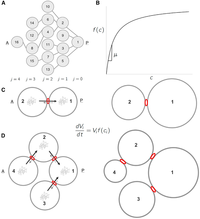
Figure 3. A Biophysical Model for Differential Growth and Emergence of Groups.
(A) Schematic representation of the ring canal tree arranged to highlight the anterior nurse cells’ spatial organization as it relates to the posterior oocyte (A, anterior; P, posterior). Each cell (node) is numbered (1–16), and each layer j is labeled (j = 0, 1…, 4). (B) A plot of the specific growth rate, f(c), as a function of concentration, c, showing both the linear regime where f(c) ≈ μc, and the saturated regime.(C) Schematic representation of a 2-cell cluster whose cells are connected by a ring canal. Each cell i of volume Vi grows according to the growth law shown. Diffusing particles of concentration, ci, in the cells’ cytoplasm that arrive at the ring canal have a higher probability of being transported from an anterior (A) cell to the more posterior (P) cell than in the opposite direction. The result is that two cells, initially of uniform size, will grow at unequal rates, with cell 1 (posterior) getting larger than cell 2 (anterior). (D) Schematic representation of a 4-cell cluster whose cells are connected by ring canals. Again, allowing for polarized transport in the posterior direction, the 4-cell cluster model leads both to differential growth—with cell 1 being largest; followed by cells 2 and 3; and, finally, by cell 4—and to the emergence of 3 groups of cell sizes that correlate with the spatial arrangement of the layers relative to cell 1.

