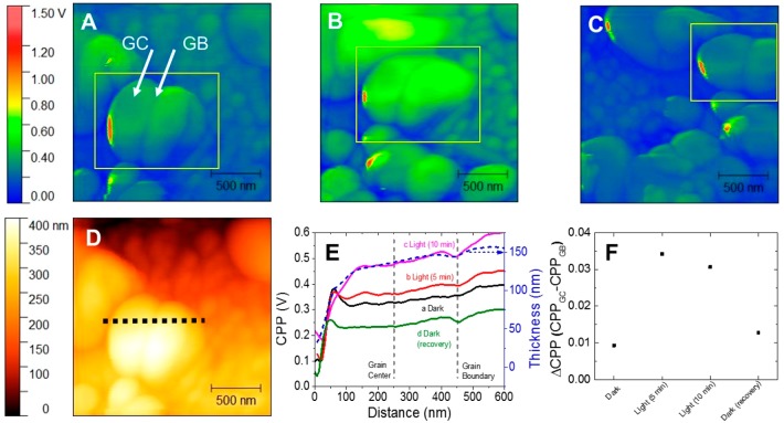
Figure 4.
Kevin probe force microscopy (KPFM) images obtained for CsPbBr3–xIx bulk films (x = 0.63) with 122 ± 27 nm grain size, under (A) dark conditions, (B) 10 min of photoirradiation, and (C) after recovery conditions in the dark. (D) Topographic image and (E) cross-sectional profile of contact point potential (CPP) through the dotted line in panel D for the bulk film. (F) CPP difference (ΔCPP) between the grain center (GC) and grain boundary (GB) in the bulk film during the dark/illumination/recovery processes. The positions of grain boundary and grain center were marked in panel E as dashed lines.

