. 2018 Oct 10;10(1):326–332. doi: 10.1039/c8sc03751e

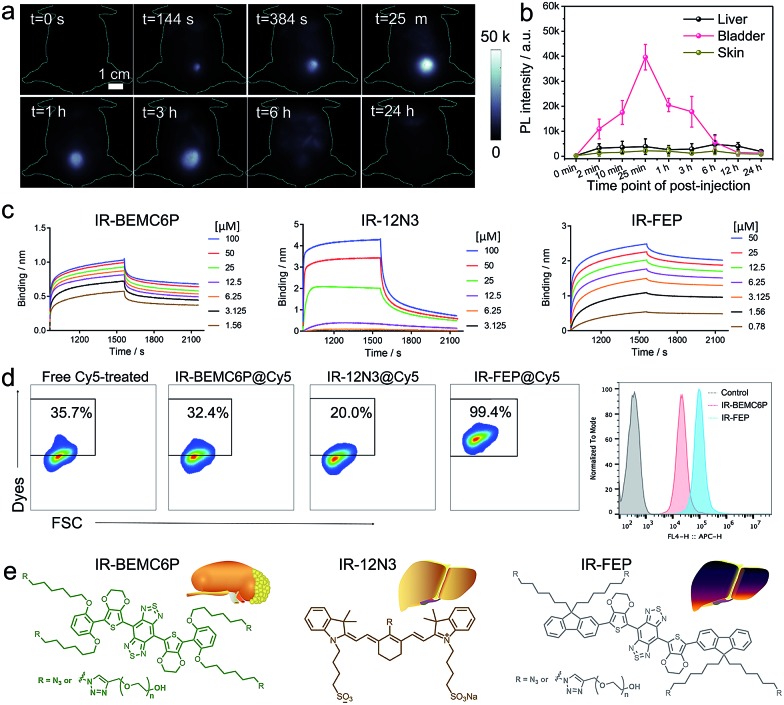
Fig. 2. The interaction between the NIR-II fluorophores and plasma protein, innate immune cells. (a) NIR-II imaging of the IR-BEMC6P injected mouse showed high bladder fluorescence signals at different p.i. time points. Injected dose: 1 mg kg–1. Imaging details: 1100 nm long pass filter, 808 nm laser. (b) Representative fluorescence signal intensity of the liver, bladder, and skin regions for IR-BEMC6P. (c) Kinetic binding assay of IR-BEMC6P, IR-12N3, and IR-FEP to albumin was measured by bio-layer interferometry. Long liver uptake dye (IR-FEP) has slower dissociating speed than short liver uptake dye (IR-12N3) and renal excretion dye (IR-BEMC6P). (d) Flow cytometry result of Cy5 labeled IR-BEMC6P, IR-12N3, and IR-FEP uptake by macrophage cells. IR-12N3 and IR-BEMC6P have much lower macrophage uptake than IR-FEP. (e) The chemical structure of renalexcretion and liver-uptake NIR-II fluorophores.

Fig. 2