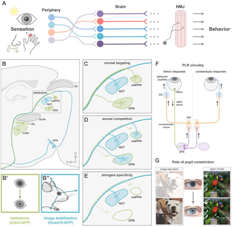
Figure 1. Retinal connections to pretectum and circuitry controlling pupillary light reflex.

(A) Sensations drive adaptive behaviors via pathways from peripheral sensory neurons to the NMJ. (B) Projection pattern of functionally distinct RGC subtypes. dLGN, dorsal lateral geniculate nucleus; IGL, intergeniculate nucleus; vLGN, ventral lateral geniculate nucleus; mdPPN, medial division of the posterior pretectal nucleus; MTN, medial terminal nucleus; NOT, nucleus of the optic tract; OPN, olivary pretectal nucleus; SC, superior colliculus. (C) Normal RGC targeting of pretectum. (D–E) Potential outcomes of losing luminance-sensing RGC input. (F) Schematic of PLR circuit. CG, ciliary ganglion; EW, Edinger-Westphal nucleus; OPN, olivary pretectal nucleus. (G) Role of pupil constriction.
