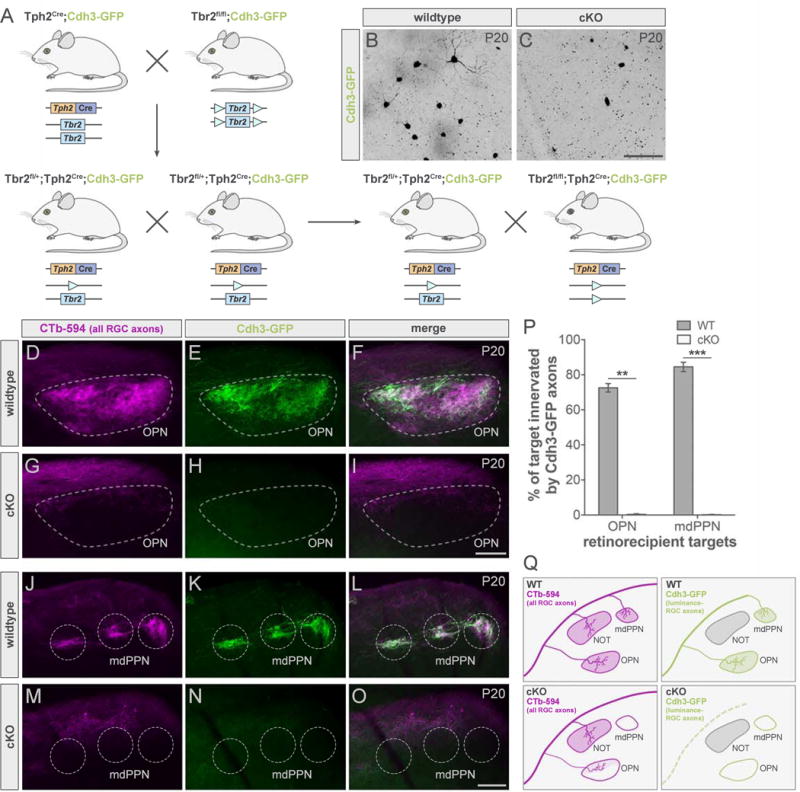
Figure 4. Conditional deletion of Tbr2 results in major loss of luminance-sensing RGCs and their axons.

(A) Floxed Tbr2 strain crossed to the Tph2Cre line to generate cKO mice (Tbr2fl/fl;Tph2Cre) mice and littermate controls (Tbr2fl/+;Tph2Cre; wildtype; WT). (B–C) Whole-mount retinas from WT (B) and cKO (C) mice with Cdh3-RGCs. Scale bar, 100 μm. (D–O) All RGC (CTb-594, magenta) and luminance-sensing RGC (Cdh3-GFP, green) axons in pretectum of WT (top panels) and cKO (bottom panels) mice. Scale bars, 100 μm. (P) Quantification of % of target innervated by Cdh3-GFP axons in WTs (dark gray) and cKOs (light gray). Data are represented as mean ± SEM (n = 3 mice per group); **p < 0.005, ***p < 0.001, Student’s t-test. (Q) Summary schematic. mdPPN, medial division of the posterior pretectal nucleus; OPN, olivary pretectal nucleus. See also Figure S3.
