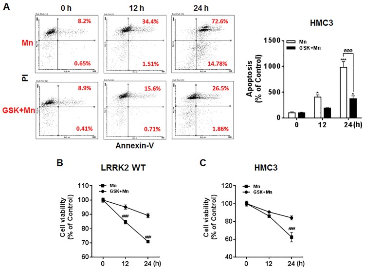
Fig 3. Inhibition of LRRK2 kinase activity attenuates Mn-induced apoptosis.
(A) After pre-treatment with GSK (1 μM) for 90 min, cells (HMC3) were exposed to Mn (250 μM) for the designated time periods, followed by annexin V and PI staining and flow cytometry analysis to determine apoptosis. Early and late apoptotic cells (Q2 and Q3) were analyzed. (B,C) After pre-treatment with GSK (1 μM) for 90 min, cells (LRRK2 WT RAW 264.7 and HMC3) were exposed to Mn for designated time periods, followed by the MTT assay to determine cell viability, as described in the Methods section, (@@@, p < 0.001; *, p < 0.05; ***, p < 0.001 compared to the control (one-way ANOVA followed by Tukey’s post hoc test; n = 3, apoptosis assay; n = 6, MTT assay). The data shown are representative of 3 independent experiments.

