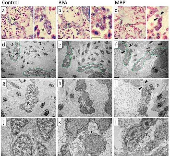
Figure 2.

Images of atrio-ventricular (AV) valve leaflets from high-level BPA and MBP exposure treatments versus solvent controls at 15 days post fertilization (dpf) Bright field images a-c are shown at ×100 magnification: AV valve leaflets were bent in the high-level MBP exposure. TEM images d-l are shown at ×3000, ×6000, and ×20 000 magnification: The extra-cellular matrix between the bilayer of valvular cells was narrower and lacked collagen in the high-level BPA and MBP exposure treatments compared to solvent controls (qualitative assessment). Annotations: A = atrium, V = ventricle, E = erythrocytes, Va = valve leaflet, IS = interstitial space, CF = collagen fibers, N = nucleus, arrow heads indicate bent AV valve leaflet. Scale bar (bottom left in each image): a–c = 10 μm; d–f = 5 μm; g–i = 2 μm; j–l = 1 μm.
