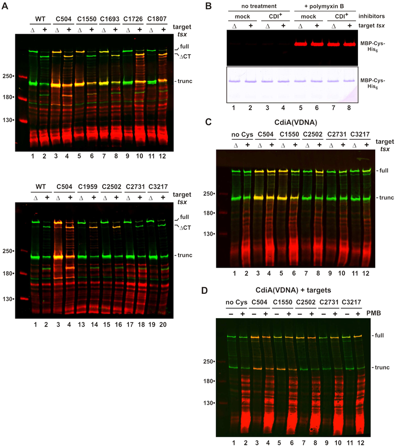
Figure 4. CdiA export resumes upon binding receptor.
A) Topology of receptor-bound CdiASTECO31. CdiA expressing cells were mixed with tsx+ or Δtsx targets and incubated with maleimide-dye. CdiA was analyzed by immunoblotting with anti-TPS antibodies. Full-length, CdiA-CT processed (ΔCT) and truncated proteins are indicated. B) Inhibitors co-expressing MBP-Cys-His6 were mixed with targets and incubated with maleimide-dye. Purified MBP was analyzed by fluorimetry (top panel) and SDS-PAGE (bottom panel). C) CdiA(VDNA) expressing cells were mixed with targets and analyzed as in panel A. D) CdiA(VDNA) expressing cells were mixed with tsx+ targets and incubated with maleimide-dye. Mixed cell suspensions were treated with PMB where indicated.

