Figure 1.

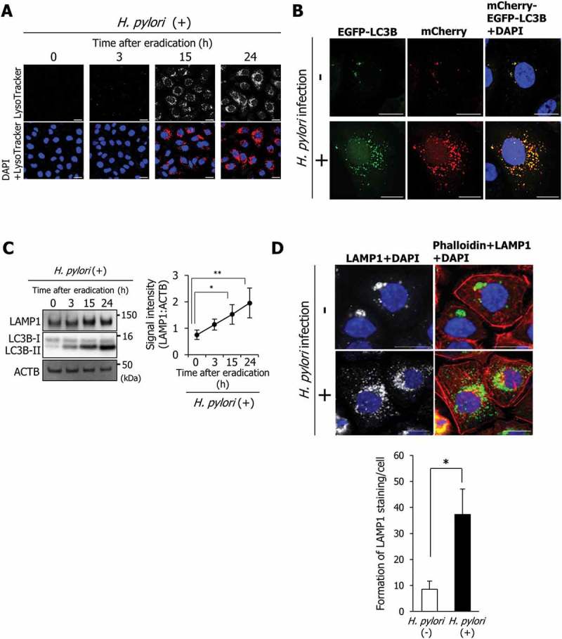
LAMP1 expression is induced during autolysosome formation. (a) AGS cells were incubated with a medium containing antibiotic for the indicated time period, infected with H. pylori for 5 h at a multiplicity of infection value of 50 (MOI 50), and stained with LysoTracker Red DND-99. Nuclei (blue) were stained with 4ʹ,6-diamidino-2-phenylindole (DAPI). Scale bar: 20 μm. (b) AGS cells were transfected with pTet-On and TRE2hyg-mCherry-EGFP-LC3B plasmids, infected with H. pylori for 5 h (MOI 50), and incubated in a medium containing antibiotic for 24 h. Then, EGFP and mCherry signals were detected. Nuclei (blue) were stained with DAPI. Scale bar: 20 μm. (C) LAMP1 levels were determined in AGS cells that were incubated with a medium containing antibiotic for the indicated duration after H. pylori infection for 5 h (MOI 50). Data are presented as the mean ± SD of 3 independent assays. *P < 0.05, **P < 0.01. (D) AGS cells were infected with H. pylori for 5 h (MOI 50) and incubated in a medium containing antibiotic for 24 h. Then, staining for LAMP1 and phalloidin staining were performed. Nuclei (blue) were stained with DAPI. Scale bar: 20 μm. The number of LAMP1-staining puncta were counted by using the ImageJ program. Data are presented as the mean ± SD of 3 independent images. *P < 0.05.
