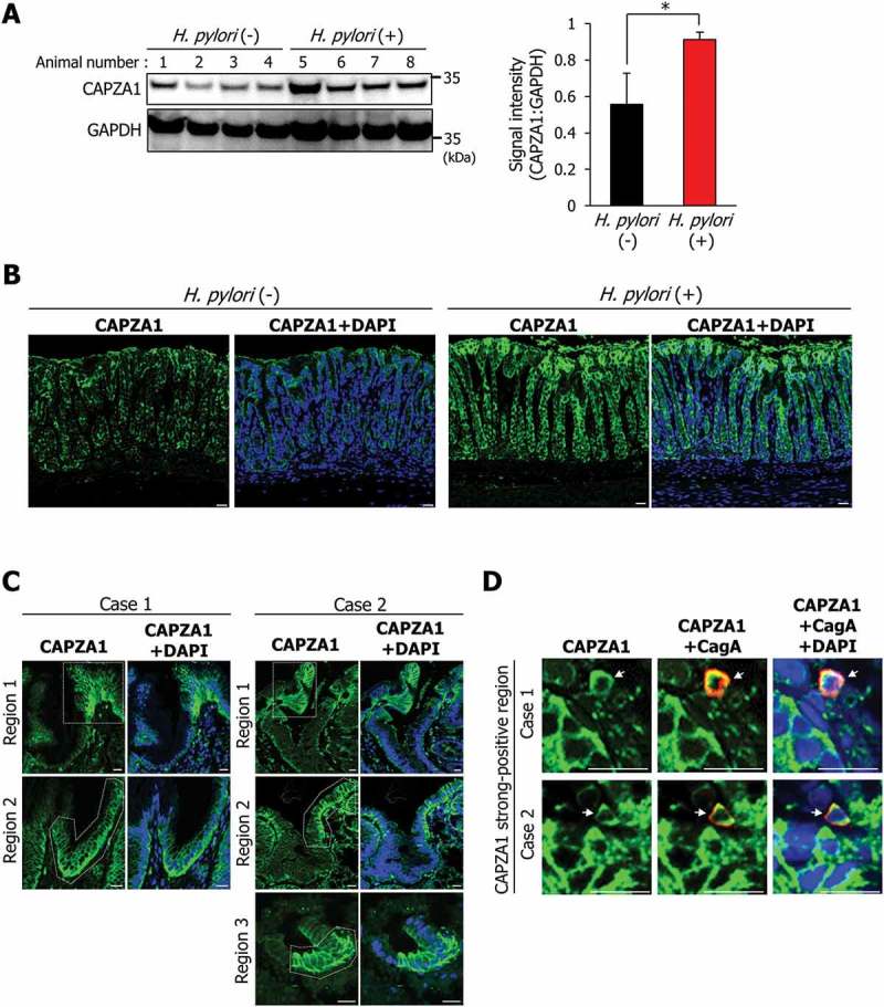
Figure 7.

CAPZA1-overexpressing cells in H. pylori-infected gastric mucosa and early gastric cancer tissues. (a) Mongolian gerbils were inoculated with H. pylori (109 CFU/mL) or vehicle. Twelve weeks after the inoculation, the animals were sacrificed, and their stomachs were excised. Western blotting using protein extracts for detection of CAPZA1 was performed. CAPZA1 signal intensity of each mouse were analyzed using the ImageJ program. *P < 0.05. (b) Staining of the stomach tissue for CAPZA1 was performed. Scale bar: 20 μm. (c) Immunostaining for CAPZA1 in human gastric adenocarcinoma. Case 1 and case 2 indicate individual gastric adenocarcinoma tissue specimens from 2 different patients. Green color indicates CAPZA1 staining. Nuclei (blue color) were stained with DAPI. The region surrounded by the white dotted line indicates a CAPZA1 strongly stained cell area. Scale bar: 20 μm. (d) Immunostaining for CagA and CAPZA1 in human gastric adenocarcinoma. Red staining indicates intracellular CagA, and green staining indicates CAPZA1. Nuclei (blue) were stained with DAPI. Scale bar: 20 μm. Arrows indicate accumulation of translocated CagA in the gastric epithelial cells strongly stained for CAPZA1.
