
Figure 2: Maternal vital signs during each episode of gastric infusion under anesthesia in “Control” group without prior anesthesia (Control).
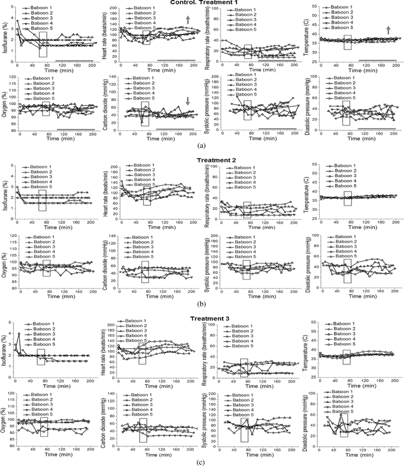
Here and in Figure 3, treatments 1, 2, and 3 correspond to gastric infusion of control (isocaloric to alcohol) solution at 90 dGa, 100 dGa, and 110 dGa, respectively. Maternal heart rate (beats/min), respiratory rate (breaths/min), peripheral capillary oxygen saturation (%), end-tidal carbon dioxide (mmHg), systolic and diastolic blood pressure (mmHg), and temperature (°C) were recorded every 10 min throughout each infusion episode. Here, and in Figures 3–5, boxed areas correspond to the time of drink infusion. Bold grey horizontal lines highlight the anesthesia time interval during which maternal vital signs were analyzed for the purpose of this retrospective study. This interval corresponds to the timing of steady maternal BAC at ≈80 mg/dL [18]. Grey arrows indicate direction of the statistically significant change in the measured parameter over the course (100–200 min) of anesthesia during treatment 1 within each group. We did not pursue the analysis during treatments 2 and 3, as fetal losses occurred after treatment 1 (Figures 3–5). For differences between the groups, please refer to Table 1. For details of statistical analysis, please refer to Section 2.5.