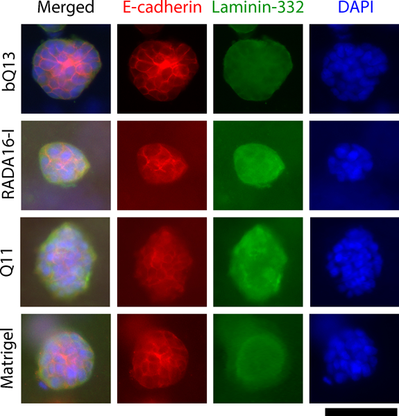
Figure 5.

Immunofluorescence of spheroids collected from dissociated matrices. E-cadherin (located at adherens junctions) and laminin-332 (secreted extracellular matrix) indicated that LNCaP spheroids exhibited an unpolarized, disorganized structure in bQ13, Q11, Matrigel, and RADA16-I. Scale bar = 100 μm.
