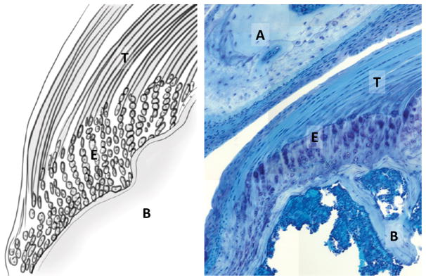
Figure 1.
Anatomy and cell morphology of the supraspinatus tendon enthesis. (Left) Schematic of the tendon-to-bone (T/B) insertion via fibrocartilage enthesis gradient. (Right) Toludine blue staining of the supraspinatus tendon inserting into the humeral head, beneath the acromion (plastic section, 20X). T: supraspinatus tendon, B: humeral bone; E: enthesis; A: acromion.

