Fig. 1.
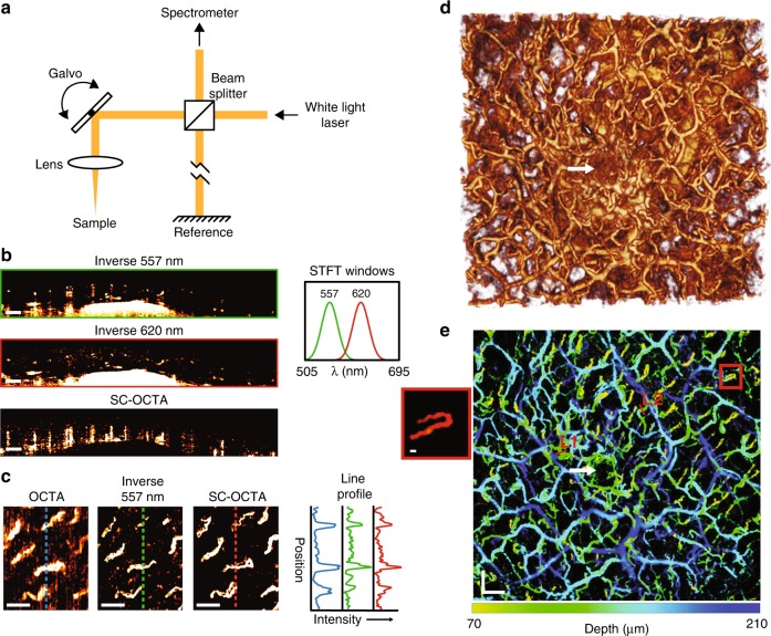
In Vivo human imaging of labial mucosa. a Simplified schematic of the visible OCT system, which allowed 3D spectral information of the sample to be obtained. b–e In vivo human imaging of labial mucosa (lower lip) from a healthy volunteer. b Inverse 557 nm and inverse 620 nm B-scans with their corresponding STFT windows, as well as a SC-OCTA B-scan showing contrast shadows from each vessel. Scale bar: 200 µm. c Comparison of angiography en face projections of superficial capillary loops with traditional motion contrast OCTA (64–111 µm), inverse 557 nm (55.6–140 µm), and SC-OCTA (83–209 µm) with their corresponding line profile intensities. Depth ranges were chosen to maximize the contrast of the en face projections for different techniques. Scale bar: 200 µm. d 3D rendering of inverse 557 nm. Scan area: 3.65 × 3.44 mm. e Depth-encoded vessel map of the same FOV as d with saturation and value from SC-OCTA and hue from the depth of the vessel in inverse 557 nm. Line 1 (L1) and Line 2 (L2) are line profiles in Figure S3 for comparison with the simulated line profile results. Scale bars: 300 µm. The red box shows a blow-up of the capillary loop; scale bar 20 µm. d, e The white arrow shows a salivary duct that is correctly not identified by SC-OCTA in e

