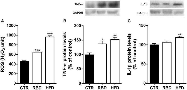
Figure 3.
Effect of regional basic (RBD) and high-fat (HFD) diets on hippocampal neuroinflammatory response. Both diets induced an (A) oxidative stress observed by an increase in reactive oxygen species (ROS) and (B) an increase of TNF-α protein levels in mice hippocampal homogenates. (C) Only HFD feeding animals show a significant upregulation of IL-1β protein levels. Above the bars, representative western blot images of TNF-α (19 kDa), IL-1β (17 kDa) and GAPDH (37 kDa) are shown. Data were analyzed by using Kruskal-Wallis test, followed by Dunn's post-test for multiple comparisons, n = 5–7. *P < 0.05, **P < 0.01, and ***P < 0.001 vs. nourished control group (CTR). The results are shown as mean ± SEM.

