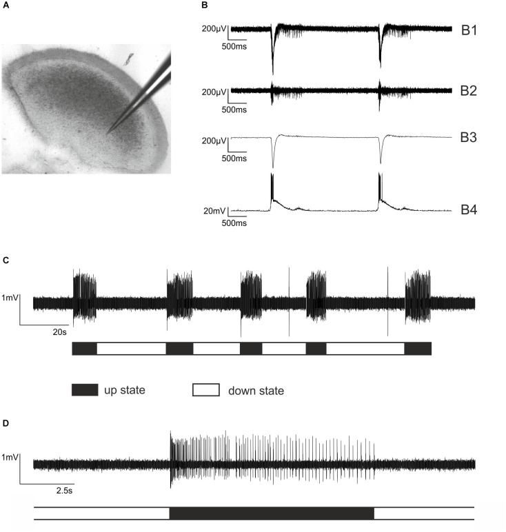
FIGURE 1.
Extracellular recordings from cortical slice cultures and firing patterns of cultured cortical neurons. (A) An electrode made of borosilicate glass placed in an organotypic cortical slice culture. Note that the cortical architecture is largely preserved. (B) Simultaneous extra- and intracellular recording from a neocortical cultured slice to illustrate the functional properties of the network. (B1) Raw trace of extracellular recording. (B2) The signal was band pass filtered 250 – 5000 Hz to allow for action potential detection. (B3) Local field potential, sometimes referred to as “micro-EEG,” which is obtained from B1 by low pass filtering 1 – 40 Hz. (B4) Simultaneous intracellular current clamp recording from a cortical pyramidal neuron within the network. The liquid junction potential of –17 mV was subtracted. (C) The typical firing pattern of cultured cortical neurons is characterized by alternating phases of high neuronal activity, so-called bursts or up states and phases of very low activity, called silent period or down state. Here, five bursts of action potential firing (up states) are displayed, halted by silent periods (down states). (D) One single up state at a higher temporal resolution. Each vertical line represents a single action potential. Note that the phase of neuronal activity at the beginning of the up state is rather high, followed by moderate activity that is slowly declining.

