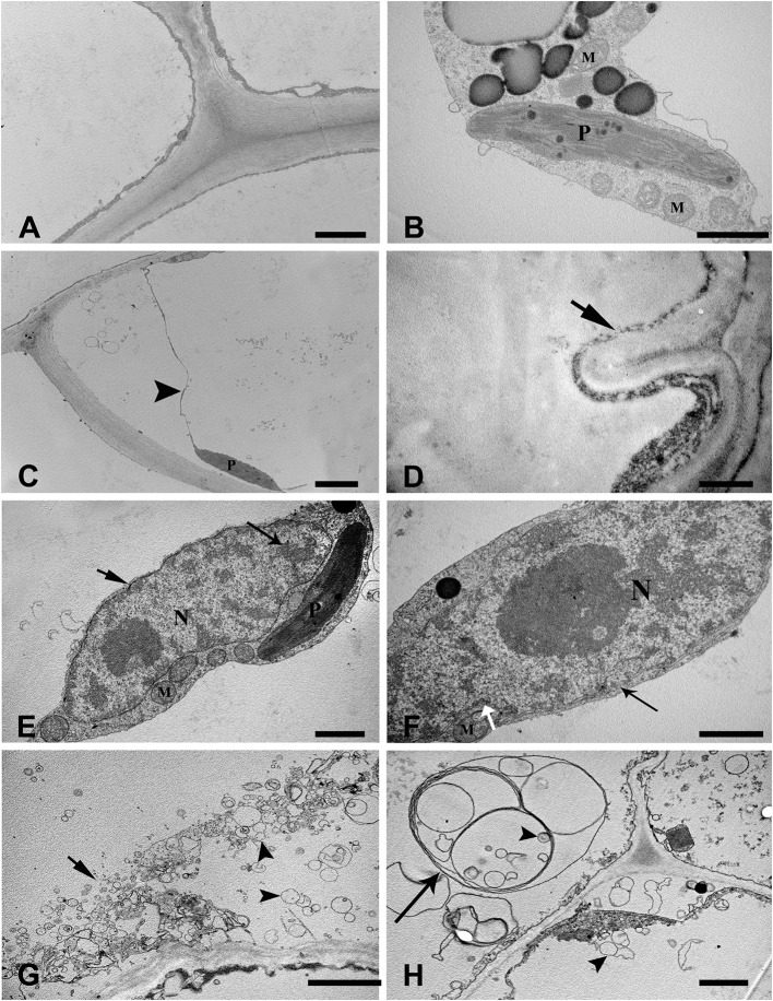
Figure 2.
Ultrastructure of aerenchyma during the early phase of formation in H. annuus stems. (A,B) Ultrastructure of the control, without WA treatment. (A) Normal ultrastructure with intact cell walls, cytoplasm and large central vacuoles. (B) Intact organelles in the cytoplasm, including mitochondria and plasmids. (C,D) Cortical cells at the early pre-cavity stage displayed plasmolysis (arrowhead) (C) and cell wall infolding (arrow) (D) after 12 h of WA treatment. (E) At 1 days after WA, condensed chromatin appeared in the nuclei (long arrow), and the nuclear envelope was heavily stained (arrow). (F) Hallmarks of nuclei degradation with nuclear invaginations (white arrow). (G) After 2 days of WA, tonoplast rupture (arrow) and lots of vesicles appearance (arrowhead). (H) Multilamellar structures (long arrow), in which some enveloped vesicles (arrowhead) were present in the cytoplasm. There are three different pictures was analyzed for each state. Bar: (A,C,G) = 2 μm, (B,E,F,H) = 1 μm, the others = 0.5 μm.

