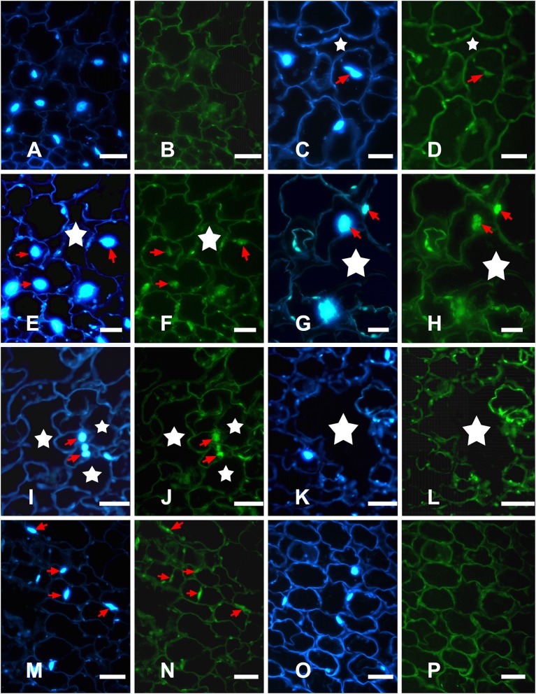
Figure 5.
Detection of in situ DNA fragmentation in H. annuus stems using DAPI staining and TUNEL assays. (A,C,E,G,I,K,M,O) DAPI assays for nuclear changes; (B,D,F,H,J,L,N,P) TUNEL assays for nuclear DNA fragmentation. (A,B) Control plants showing no aerenchyma and DAPI-positive and TUNEL-negative staining. (C,D) In early phase of aerenchyma formation (star), TUNEL-positive nuclei were first detected in certain cells of the cortex. (E,F) In the formation phase of aerenchyma formation (star), TUNEL-positive nuclei were observed around the intercellular space; (G–J) In the expansion phase of aerenchyma formation (star), the remaining cells surrounding the lacunae showed a high frequency of TUNEL-positive nuclei. (K,L) In the mature phase of aerenchyma formation (star), no TUNEL-positive nuclei were observed in the stem tissues; (M,N) TUNEL-positive control; (O,P) TUNEL-negative control.

