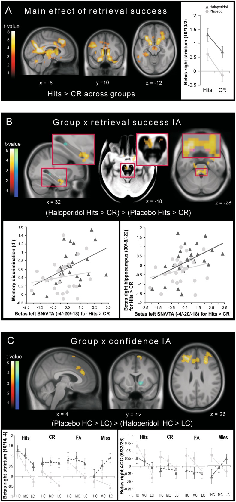
Fig. 3.

fMRI results. a Activity pattern of retrieval success across groups and β estimates from the peak voxel in the right striatum. b Significantly increased activity for retrieval success under haloperidol in the hippocampus and amygdala (left), SN/VTA (middle; activation displayed on the mean MT image showing the SN/VTA as a bright region) and LC (right; activation overlaid with the LC mask [73] in magenta). Scatter plots of the individual SN/VTA response for retrieval success and memory accuracy (left) and the individual anterior hippocampus response for retrieval success (right) in the haloperidol (dark gray triangles) and the placebo group (light gray circles). c Regions showing higher confidence activity under placebo and β plots illustrating the confidence pattern in the right striatum and the ACC. Dashed lines represent the linear trend across confidence levels. All activation maps are thresholded at p < 0.05 (warm colors: FWE-corrected at the cluster level using a cluster forming threshold at voxel level of p < 0.001; cold colors: small-volume FWE-corrected using anatomical masks). The inverse contrasts revealed no significant activations. HC high confidence, MC medium confidence, LC low confidence, IA interaction
