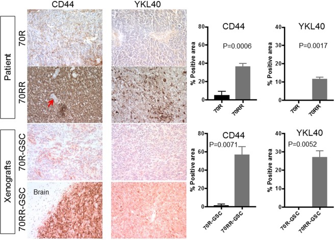
Figure 6.
MGG70RR tumour and its GSC xenografts overexpress mesenchymal markers. Immunohistochemistry (IHC) for CD44 and YKL-40 in the patient tumours, MGG70R (70R) and MGG70RR (70RR), and the corresponding orthotopic GSC xenografts. Upper two rows, patient tumours. Lower two rows, xenografts. For CD44 IHC in MGG70RR-GSC tumours, a tumour-brain border area was depicted to demonstrate tumour-selective staining. Note negative staining of CD44 in tumour-associated blood vessels (arrow). Image quantification of IHC is shown on the right: upper plots, patient tumors; lower plots, xenografts.

