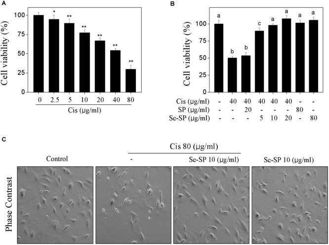
FIGURE 2.

Selenium-SP inhibits cisplatin-induced cytotoxicity in MC3T3-E1 cells. (A) Dose-dependent cytotoxicity of cisplatin on MC3T3-E1 cells. Cells seeded in 96-well plate were treated with 0–80 μg/ml cisplatin for 24 h. (B) Se-SP inhibited cisplatin-induced cytotoxicity in MC3T3-E1 cells. Cells seeded in 96-well plate were pre-treated with 5–20 μg/ml SP or Se-SP for 24 h, and co-treated with 40 μg/ml cisplatin for another 24 h. Cell viability was detected by MTT assay. (C) Phase contrast of MC3T3-E1 cells morphology. Cells morphology was examined by phase-contrast microscopy. All data and images were obtained from three independent experiments. Bars with “∗” or “∗∗” indicates P < 0.05 or P < 0.01, respectively, when compared with control group. Bars with different characters are statistically different at P < 0.05 level, which achieved the multiple comparisons.
