Figure 2.
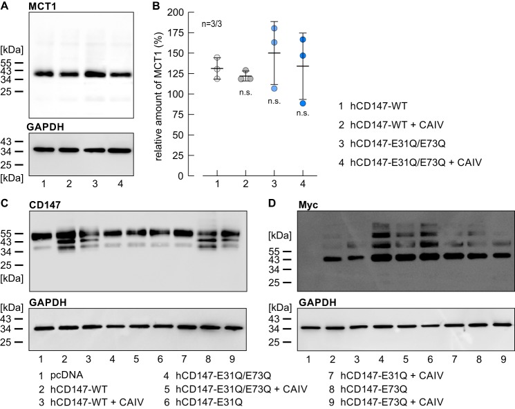
Expression of MCT1 and CD147 in HEK-293 cells. A, representative Western blotting against MCT1 (upper blot) and GAPDH (lower blot) as loading control, from HEK-293 cells transfected with human CD147–WT (lane 1), hCD147–WT, and CAIV (lane 2), the mutant hCD147–E31Q/E73Q (lane 3), and hCD147–E31Q/E73Q and CAIV (lane 4), respectively. B, quantification of the relative protein level of MCT1 (as normalized to the signal of GAPDH in the same lane) in HEK-293 cells, transfected with hCD147–WT (lane 1), hCD147–WT + CAIV (lane 2), hCD147–E31Q/E73Q (lane 3), and hCD147–E31Q/E73Q + CAIV (lane 4), respectively. The significance indicators refer to the values of HEK-293 cells, transfected with hCD147–WT. n is given as the number of Western blots/number of batches of cells. C, representative Western blotting against CD147 (upper blot) and GAPDH (lower blot) as loading control, from HEK-293 cells, transfected with the empty vector pcDNA3 (lane 1), hCD147–WT or a mutant of CD147 (lanes 2, 4, 6, and 8), or cotransfected with hCD147 or a mutant of CD147 and CAIV (lanes 3, 5, 7, and 9). D, representative Western blotting against the Myc tag of the transfected CD147 (upper blot) and GAPDH (lower blot) as loading control, from HEK-293 cells, transfected with the empty vector pcDNA3.1 (lane 1), hCD147–WT or a mutant of hCD147 (lanes 2, 4, 6, and 8), or cotransfected with hCD147 or a mutant of hCD147 and CAIV (lanes 3, 5, 7, and 9).

