Figure 3.

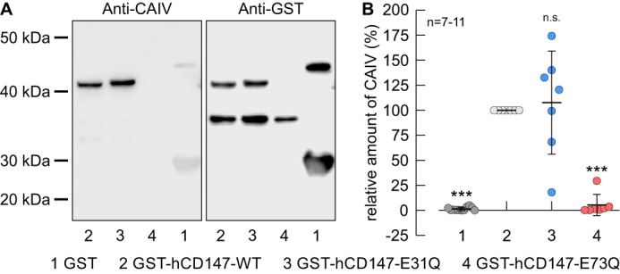
Binding of CAIV to hCD147 requires the Glu-73 in the Ig1 domain of hCD147. A, representative Western blots of CAIV (left blot) and GST (right blot), respectively. CAIV was pulled down with GST (lane 1), a GST fusion protein of the Ig1 domain of hCD147–WT (lane 2), a GST fusion protein of Ig1 domain of the hCD147 mutant E31Q (lane 3), and a GST fusion protein of Ig1 domain of the hCD147 mutant E73Q (lane 4). B, relative intensity of the fluorescent signal of CAIV. For every blot, the signals for CAIV were normalized to the corresponding signals for GST–hCD147–WT. Each individual signal for CAIV was normalized to the intensity of the signal for GST in the same lane. The significance indicators above the dots refer to GST–hCD147–WT.
