Figure 1.
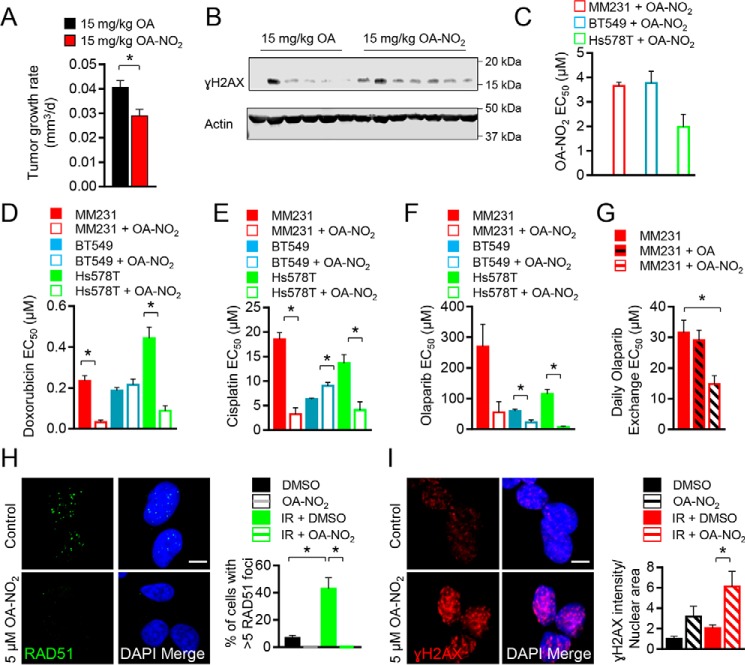
OA-NO2 inhibits TNBC cell growth, RAD51 foci formation, and sensitivity to ionizing radiation. A, MDA-MB-231 (MM231) cells (0.5 × 10⁶) were orthotopically injected into 6-week-old mice, and mice were gavaged with 15 mg/kg OA (black) or OA-NO2 (red) for 4 weeks when tumors reached a volume of 100 mm3. Values indicate average, and error bars represent S.E.; n = 6–7 per group. B, tumoral γH2AX was increased in OA-NO2–treated mice compared with OA control mice as assessed by immunoblotting (n = 6–7 per group). C, MDA-MB-231 (red), BT549 (blue), or Hs578T (green) cells were treated with increasing concentrations of OA-NO2, and relative growth was measured by quantifying luminescent ATP levels (CellTiter-Glo). EC50 values indicate average, and error bars represent S.E.; n = 3. D–F, MDA-MB-231 (red), BT549 (blue), or Hs578T (green) cells were treated with increasing concentrations of doxorubicin, cisplatin, or olaparib ± OA-NO2 and measured as above. G, MDA-MB-231 (red) cells were treated with increasing concentrations of olaparib daily + vehicle, OA, or OA-NO2 and measured as above. H and I, OA-NO2 diminished RAD51 foci formation (green) and increased γH2AX (red) in MDA-MB-231 cells following irradiation with 5 Gy. Merged samples include 4′,6-diamidino-2-phenylindole (DAPI)-stained nuclei (blue). Cells on 16-well coverslips were dosed with 5 Gy and then treated with 5 μm OA-NO2 or vehicle for 6 h prior to immunofluorescence processing. The average percentages of cells with five or more foci from confocal z-stacked images are indicated from 0- or 5-Gy samples, and error bars represent. * = p < 0.05.

