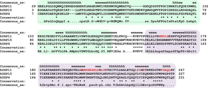
Figure 3.
Sequence alignment of USP11, USP4, and USP15 DU domains. Shown is structure-based sequence alignment using PROMALS3D (61) of human USP11 (PDB code 4MEL (20)), USP15 (PDB code 3T9L (21)), and USP4 (PDB code 3JYU, Structural Genomics Consortium (SGC), J. P. Bacik, G. Avvakumov, J. R. Walker, S. Xue, and S. Dhe-Paganon, unpublished data) with secondary structure elements above the sequences indicated (e = strand; h = helix). The DUSP domain is shaded green, and the UBL domain is shaded purple. Sequence conservation is depicted as per PROMALS3D default representation (bold uppercase letters (such as G); aliphatic residues (I, V, L): 1, aromatic residues (Y, H, W, F); @, hydrophobic residues (W, F, Y, M, L, I, V, A, C, T, H); h, alcohol residues (S, T); o, polar residues (D, E, H, K, N, Q, R, S, T); p, tiny residues (A, G, C, S); t, small residues (A, G, C, S, V, N, D, T, P); s, bulky residues (E, F, I, K, L, M, Q, R, W, Y), b, positively charged residues (K, R, H); +, negatively charged residues (D, E); −, charged (D, E, K, R, H) with the exception that identical residues are indicated using an asterisk. USP11 UBL domain residues located at the interface upon peptide binding are highlighted in red. aa, amino acid.

