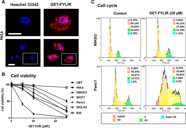
Figure 6.
Cellular effects of FYLIR peptide agent. A, GET-FYLIR has cytoplasmic, nuclear, and nucleolar localization. HeLa cells were treated with GET-FYLIR (10 μm) for 24 h and assessed by confocal microscopy (nuclei were counterstained with Hoechst 33342). The white scale bar is 20 μm. B, GET-FYLIR decreases cell viability cell type–specifically and dose-dependently. Cell viability (Presto Blue assay) was assessed after 24 h of incubation with GET-FYLIR (0, 10, 20, and 50 μm) in KNS-42, U87, BJ6, HeLa, NIH3T3, MCF7, and Panc1 cells. Cell viability was expressed as percentage of cell viability ± S.D. (n = 3 biological repeats). C, GET-FYLIR induces S phase arrest cell type–specifically. Cell cycle analysis was conducted using NIH3T3 cells (n = 5 biological repeats) and Panc1 cells (n = 7 biological repeats) untreated or incubated with GET-FYLIR (20 μm) for 24 h. Cells were fixed, stained with propidium iodide staining solution, and analyzed for DNA content. The distribution and percentage of cells in subG0, G0, S, G2, and super-G2 phase of the cell cycle are indicated.

