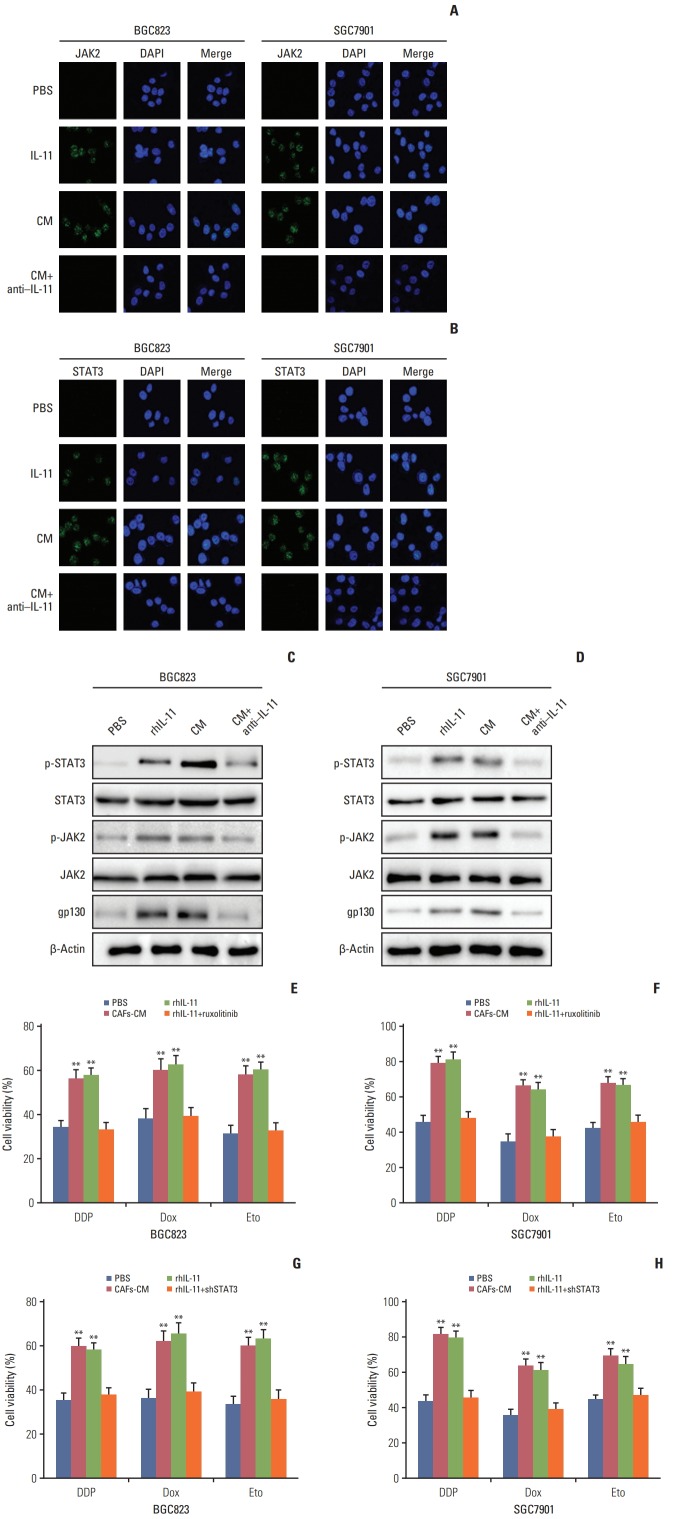
Fig. 3.
Interleukin 11 (IL-11)/IL-11R signaling pathway induced the chemo-resistance through JAK/STAT3 pathway. (A) Immunofluoresence of p-JAK2 in BGC823 and SGC7901 cells pre-treated with or without cultured medium of cancer-associated-fibroblasts (CAFs-CM)/rhIL-11 in the presence or absence of IL-11 neutralizing antibody. (B) Immunofluoresence of p-STAT3 in BGC823 and SGC7901 cells pre-treated with or without CAFs-CM/rhIL-11 (10 ng/mL) in the presence or absence of IL-11 neutralizing antibody (25 μg/mL). (C) Western blotting of p-JAK2, total JAK2 and β-actin in BGC823 and SGC7901 cells pre-treated with or without CAFs-CM/rhIL-11 (10 ng/mL) in the presence or absence of IL-11 neutralizing antibody (25 μg/mL). (D) Western blotting of p-STAT3, total STAT3, and β-actin in BGC823 and SGC7901 cells pre-treated with or without CAFs-CM/rhIL-11 (10 ng/mL) in the presence or absence of IL-11 neutralizing antibody (25 μg/mL). (E) The cell viability of BGC823 cells treated with 6 μg/mL DDP, 6 μM etoposide, and 6 μM doxorubicin respectively with or without CAFs-CM or rhIL-11 (10 ng/mL) pre-co-cultured in the presence or absence of ruxolitinib (5 μM). PBS, phosphate buffered saline; Dox, doxorubicin; Eto, etoposide. (F) The cell viability of SGC7901 cells treated with 4 μg/mL DDP, 6 μM etoposide, and 6 μM doxorubicin respectively with or without CAFs-CM or rhIL-11 (10 ng/mL) pre-co-cultured in the presence or absence of ruxolitinib (5 μM). (G) The cell viability of BGC823 cells treated with 6 μg/mL DDP, 6 μM etoposide, and 6 μM doxorubicin respectively with or without CAFs-CM or rhIL-11 (10 ng/mL) pre-co-cultured in the wild type or shSTAT3 cells. (H) The cell viability of SGC7901 cells treated with 4 μg/mL DDP, 6 μM etoposide, and 6 μM doxorubicin respectively with or without CAFs-CM or rhIL-11 (10 ng/mL) pre-co-cultured in the wild type or shSTAT3 cells. The data was presented as the mean±standard error of mean from three independent experiments. **p < 0.01.

