
Figure 1.
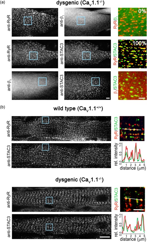
STAC3 localizes in the triads of dysgenic (CaV1.1−/−) myotubes in vitro and in vivo. (a) Representative immunofluorescence images of dysgenic myotubes stained to label the ryanodine receptor (RyR), the DHPR β1a subunit, and STAC3. Scale bar = 10 μm. The numbers indicate the percentage of myotubes in which β1a or STAC3 colocalized with the RyR (N = 3, n = 90). Color overlays are 4× magnifications of the regions indicated by the blue squares. Scale bar = 5 μm. (b) Diaphragm muscle fibers of wildtype and dygenic mice, double‐labeled with anti‐RyR and anti‐STAC3. Scale bar = 10 μm. 4× magnification color overlays of STAC3 (green) and RyR (red). Scale bar = 2.5 μm. Fluorescence intensity scans along two sarcomeres of both wildtype (upper panel) and dysgenic (lower panel) muscles show peaks for STAC3 (green) and RyR1 (red) at exactly the same location consistent with their colocalization in the triads. DHPR, dihydropyridine receptor [Color figure can be viewed at wileyonlinelibrary.com]