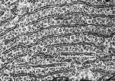
Figure 2.

Electron microscopy shows that Nissl bodies in a motor neuron are stacks of rough endoplasmic reticulum whose cisterns are studded externally with ribosomes (white rectangles) and interspersed with rosettes of polyribosomes (white circles). [Image taken from Palay in (Fawcett, 1981) (p.319) in which magnification is not stated but it was noted that fenestrated cisternae are separated by intervals of 0.2 to 0.5 μm].
