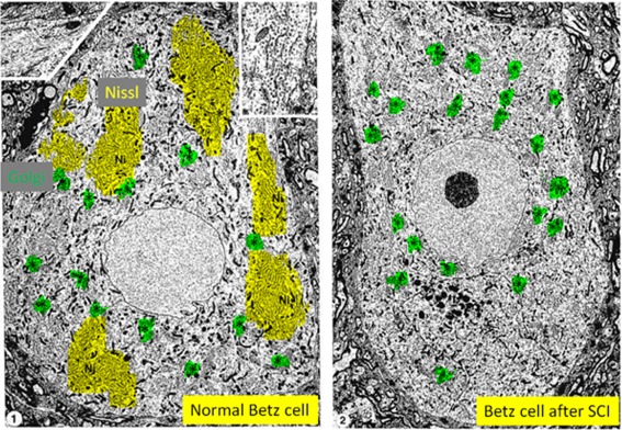
Figure 3.

Chromatolysis in CNS neurons involves destruction of the Nissl body component of the protein synthesis machinery. Electron microscope image showing Betz neurons from pericruciate cortex of either (panel 1) a normal adult cat or (panel 2) an adult cat, ten days after spinal cord injury (C2 lateral funiculotomy). Nissl substance is highlighted in yellow (Ni) and aggregates of Golgi are highlighted in green (*). Normal Nissl is no longer visible in the cortical neuron after spinal cord injury. [Images from (Barron and Dentinger, 1979); magnification of panel 1 is X 5,300 (inset is X 21,700) and magnification of panel 2 is X 3,400].
