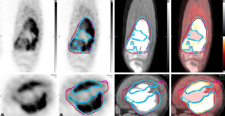
Figure 5.
MO-PET and PETedge segmentation in a patient with osteosarcoma. Repeated independent measurements using MO-PET (blue and light blue colored ROIs) and PETedge (red and pink colored ROIs) showed excellent intra-method agreements with the minor discrepancy. Note that the tumor portion with a low FDG uptake was included in PETedge segmentation but excluded in MO-PET segmentation.

