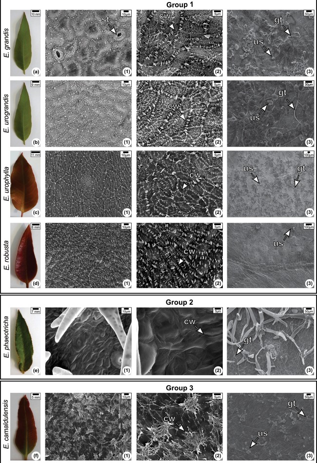
FIGURE 5.
Leaf morphology (a–f) and scanning electron micrograph of the adaxial surface of the leaves of Eucalyptus species non-inoculated (1–2) and inoculated (3) with Austropuccinia psidii at 144 h.a.i Epicuticular wax morphology: Group I—platelets; Group II—Wax sheet; Group III—Tubes or threads. (1) and (2) were observed using different magnifications (1,100× and 3,000×, respectively). CW, cuticular wax; ST, stomata; GT, germination tube; US, Uredospore.

