Fig. 1.

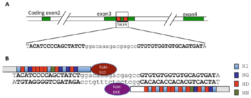
Targeting TRα with TALEN in Xenopus tropicalis embryos. (a) Schematic diagram of the TRα gene showing the TALEN target site. The protein coding exons were shown as numbered, colored boxes. The TALEN-recognized sequences were shown as red boxes (top) or bold letters (below). (b) Schematic representation of the TRα TALEN and the targeted DNA sequence in the TRα gene. The four types of RVDs (repeat variable di-residues) recognizing nucleotide A, G, T, or C were depicted in different colors, respectively. The left arm contained the FokI-ELD nuclease and the right arm contained the FokI-KKR nuclease, which, when both arms bind to their respective binding sites, heterodimerize to form the functional nuclease to make a double-stranded break in the intervening sequence. See [35] for more details
