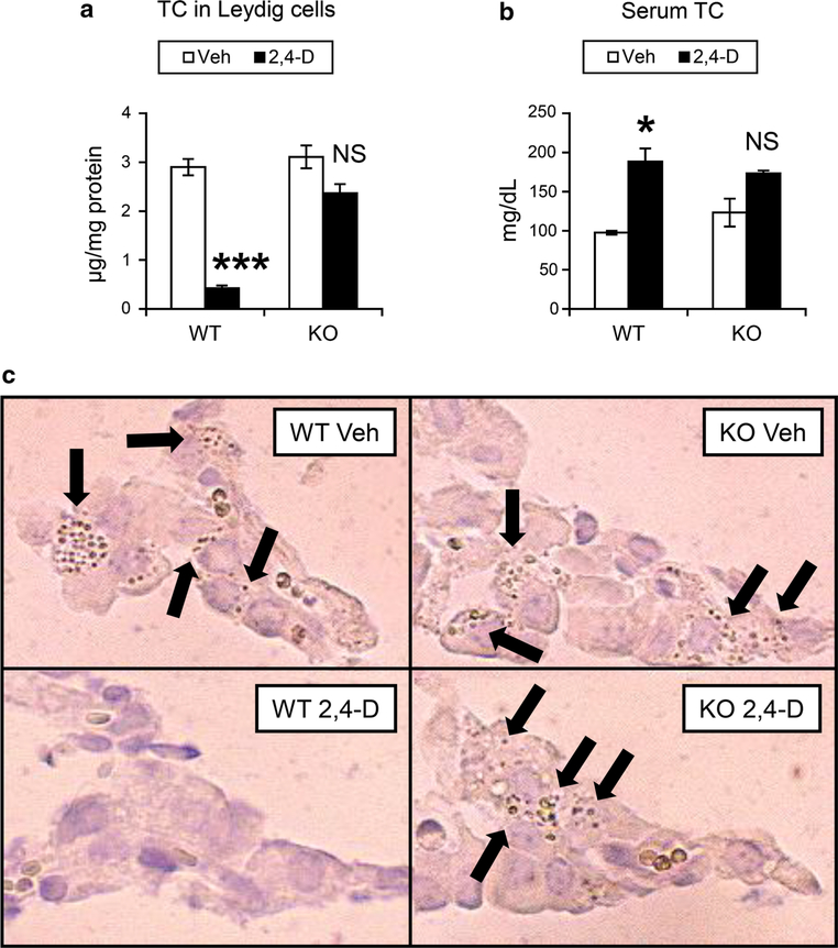
Fig. 4.
2,4-D treatment depleted cholesterol contents in Leydig cells in a PPARα-dependent manner. a, b Total cholesterol (TC) levels in isolated Leydig cells (a) and serum (b) were determined using the samples obtained from vehicle (Veh)- or 2,4-D-treated Sv/129 wild-type (WT) or Ppara-null (KO) mice. Values were expressed as mean ± SEM (n = 5). Statistical analysis was performed using ANOVA test with Bonferroni’s correction. *P < 0.05; ***P < 0.001; NS not significant between the 2,4-D-treated and Veh-treated mice in the same genotype. c The abundance of cholesterol ester in Leydig cells was assayed using frozen testis sections obtained from vehicle (Veh)- or 2,4-D-treated Sv/129 wild-type (WT) or Ppara-null (KO) mice and cytochemical staining according to the method of Emeis et al. Arrows indicate cholesterol-rich particles

