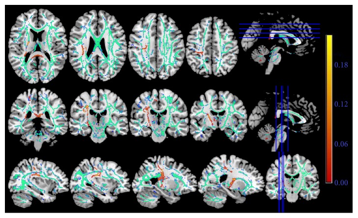
Figure 1.
The FA skeleton based on the diffusion tensor imaging. The red color indicated the injured white matter fibers in the stroke patients in contrast to the healthy controls. The green color represents the common white matter tracts between two groups of participants. The right-hemispheric subcortical stroke patients displayed significantly decreased fractional anisotropy in the ipsilesional superior longitudinal fasciculus, corticospinal tract, and the corpus callosum.

