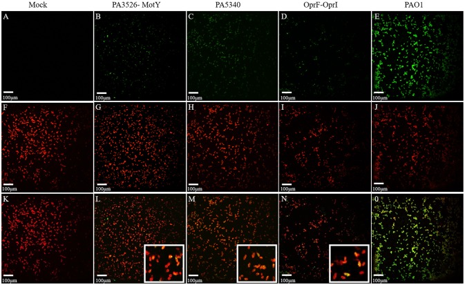
Figure 4.
Cellular localization of vaccine candidates PA5340 and PA3526-MotY and controls by immunofluorescence microscopy. Immunofluorescence staining with confocal microscopy shows the localization of antigens (green) (A–E) and the PAO1 cell wall (red) (F–J). For antigens localization the antisera of naïve mice (A) or immunized with PA3526-MotY (B), PA5340 (C), OprF-OprI (D) or heat inactivated PAO1 (E) were used. Merged images show the co-localization of the two signals (yellow) (K–O) suggesting that proteins could be surface exposed. Detailed co-localization of antigens of interest is shown in the magnification (L, M, N).

