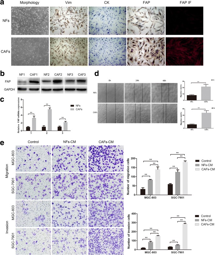
Fig. 1.
Characterization of primary cultured NFs and CAFs and their effects on migration and invasive ability of GC cells. a The morphology of gastric NFs and CAFs (left). Immunocytochemical staining showed the expression of Vimentin, Cytokeratin, and FAP in NFs and CAFs (middle), and immunofluorescence staining for FAP (right). b Western blot analysis of FAP expression in three paired NFs and CAFs. c The mRNA expression levels of FAP in three paired NFs and CAFs. d The migration ability of CAFs itself was stronger than that of paired NFs at 48 h and 72 h. e CAFs-CM significantly promoted the migration and invasive ability of MGC-803 and SGC-7901 cells than NFs-CM. (*P < 0.05, **P < 0.01)

