. 2018 Nov 15;8(1):15–24. doi: 10.1039/c8tx00231b

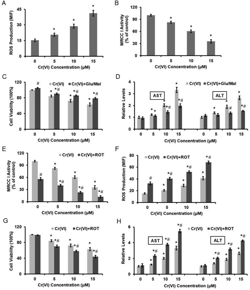
Fig. 2. Cr(vi)-Induced cytotoxicity was associated with the inhibition of MRCC I activity. L-02 hepatocytes were exposed to different concentrations of Cr(vi) (0, 5, 10, and 15 μM) for 24 h. (A) ROS level was measured using HE and was quantified by the MIF. (B) MRCC I activity was determined using the commercial kit. L-02 hepatocytes were treated with the MRCC I substrates 10 mM Glu/10 mM Mal for 10 min prior to Cr(vi) exposure. (C) Cell viability. (D) AST and ALT levels. ROT (5 μM, 24 h) was used to treat L-02 hepatocytes together with Cr(vi). (E) MRCC I activity. (F) ROS level. (G) Cell viability. (H) AST and ALT levels. *p < 0.05, compared with control. #p < 0.05, compared with Cr(vi) alone treatment.

Fig. 2