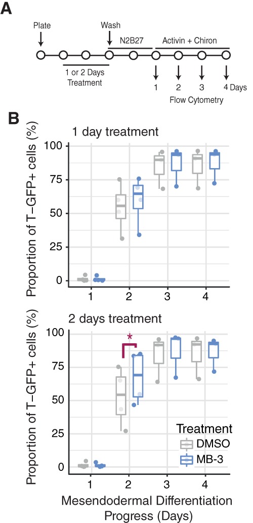
Figure 4.

Effect of MB‐3 treatment on mesendodermal differentiation of mouse embryonic stem (ES) cells. (A): Mesendodermal differentiation experimental design: Brachyury‐GFP (T‐GFP) cells treated with MB‐3 or DMSO in ESLIF medium (1–2 days) before wash‐off and replating in N2B27 (days 0–1) followed by N2B27 with 100 ng/ml Activin and 3 μM Chiron (days 2–4). Flow cytometry was used to quantify the proportion of GFP+ cells on days 1–4 of differentiation. (B): Exposure of mouse ES cells to MB‐3 for 2 days significantly anticipates detection of T‐GFP expression, indicative of accelerated ME commitment (n = 4). Paired Student's t test. *p < .05. Abbreviations: DMSO, dimethyl sulfoxide; GFP, green fluorescent protein.
