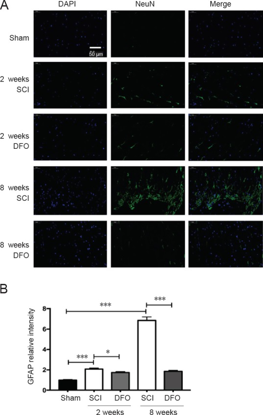
Figure 7.

Effects of DFO on GFAP immunofluorescence at 2 and 8 weeks after injury.
Immunofluorescent images of GFAP+ cells in transverse sections were captured using a fluorescence microscope at the center of injured spinal cord. Scale bar: 50 μm. (A) Representative fluorescence micrographs of GFAP (in green) staining and cell nuclei (in blue) in the injury epicenter. (B) Quantification of GFAP in the injury epicenter. *P < 0.05, ***P < 0.001. Data are expressed as the mean ± SEM (n = 3; two-way analysis of variance followed by Tukey’s post hoc test). DAPI: 4′,6-Diamidino-2-phenylindole; DFO: deferoxamine; GFAP: glial fibrillary acidic protein; SCI: spinal cord injury.
