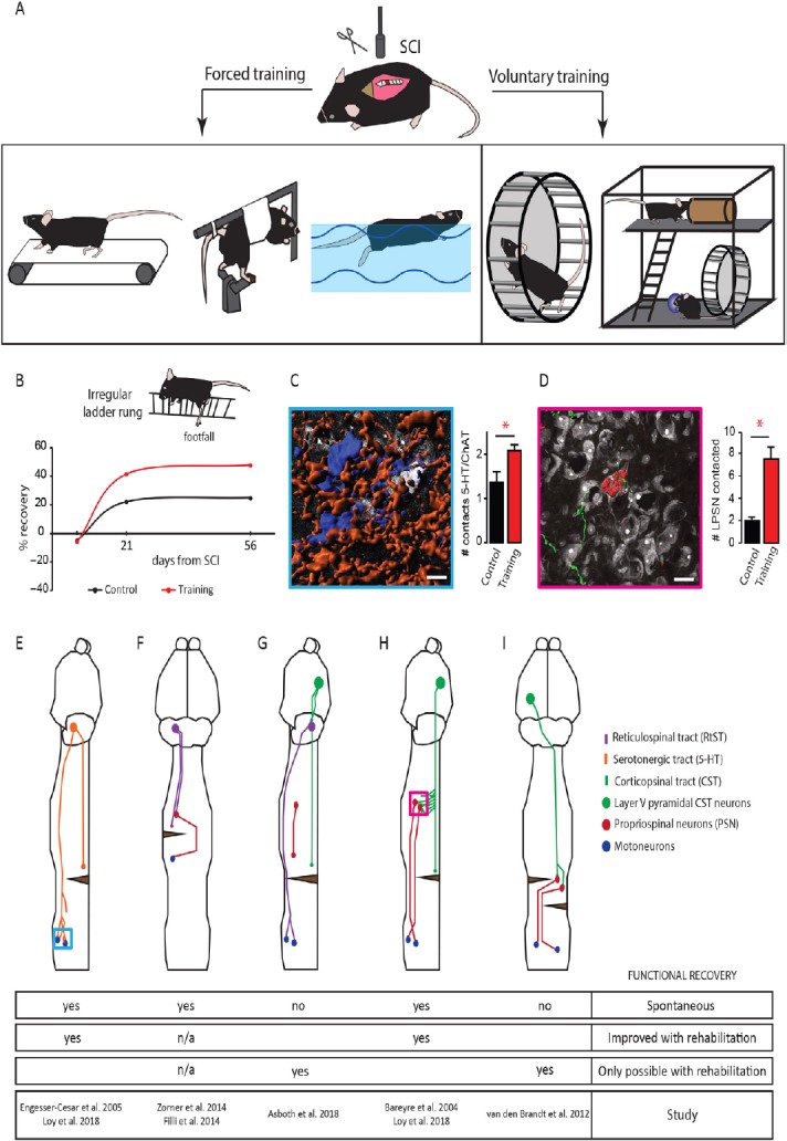
Figure 1.

Overview of animal models of spinal cord injury (SCI) rehabilitation and schematic representations of several detour circuits paradigms in SCI.
(A) Overview of several experimental approaches used to rehabilitate hindlimb function following SCI in rodents. Rehabilitation can be divided into forced approaches (from left to right): treadmill walking, passive bicycling and swimming and voluntary strategies (from left to right): wheel running and enriched environments. (B) Schematic representation of the percentage of recovery obtained in the irregular ladder rung test with voluntary wheel running. (C) Representative rendering (left) of lumbar motoneurons (blue) contacted by serotonergic fibers (orange) and quantification of the number of contacts between serotonergic (5-HT) fibers and motoneurons 2 weeks after SCI without and with training on an irregular wheel (right). Displayed in white are non-positive neurons. (D) Representative rendering (left) of a corticospinal tract collateral (green) contacting a long propriospinal neuron (red) in the cervical spinal cord 2 weeks after dorsal midthoracic hemisection and quantification of the number of long propriospinal neurons contacted 2 weeks after lesion without and with training on an irregular running wheel (right). Displayed in white are non-positive neurons labeled. Scale bars: 20 µm. *P < 0.05. # is an abbreviation for number. (E) Schematic representation of the remodeling of serotonergic fibers in the spinal cord following dorsal midthoracic hemisection. Blue inset refers to image in panel C. (F) Schematic representation of the sprouting and detour circuit formation of the reticulospinal tract following a cervical hemilesion. (G) Schematic representation of a detour circuit of the corticospinal tract via the reticulospinal tract following a near complete thoracic lesion. (H) Schematic representation of the corticospinal tract-long propriospinal neuron detour circuit following midthoracic dorsal hemisection. Pink inset refers to image in panel D. (I) Schematic representation of a corticospinal-propriospinal detour circuit following temporally and spatially separated complete lesion of the spinal cord (staggered hemisection). ChAT: Choline acetyltransferase; LPSN: left propriospinal neuron.
