
Figure 6.
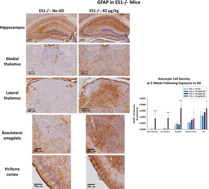
Effect of GD‐induced status epilepticus and delayed midazolam treatment on astrocytic cell response in male ES1−/− mice. At 2 weeks following exposure to GD and delayed midazolam treatment, ES1−/− mice were perfused and brains collected for immunohistochemistry processing with an antibody against GFAP, a marker for astrocytes. Cresyl violet was used as counterstain for visualization of anatomic landmarks. GFAP‐positive (GFAP+) cells were manually counted in the bregma range of −1.28 to −1.64 mm in the dorsomedial thalamus, dorsolateral thalamus, basolateral amygdala, layer 3 of the piriform cortex, and the CA1 region of the hippocampus. GFAP+ cell density is shown in graph. *P < 0.05, ***P < 0.001 compared to control (No GD) ES1−/− mice