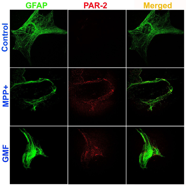
Fig. 4.
MPP+ and GMF upregulate the expression of PAR-2 in mouse primary astrocytes as determined by double immunofluorescence staining. Astrocytes were incubated with MPP+ (10 μM) or GMF (100 ng/ml) for 48 hrs and the expression of PAR-2 and GFAP were analyzed by immunofluorescence staining (n=3). The cells were incubated with monoclonal antibody for PAR-2 and polyclonal primary antibodies for GFAP followed by incubation with Alexa Flour 488 goat anti-rabbit IgG and Alexa Flour 568 goat anti-mouse IgG secondary antibodies. Then the cover glass with cells were lifted from the wells and mounted on to the microscope glass slides, dried and viewed using a confocal microscope. Representative images show that astrocytes incubated with MPP+ and GMF increased the expression of PAR-2 (red fluorescence) as compared with untreated control cells. Astrocyte marker GFAP is shown by the green fluorescence. Merged images show co-localization of PAR-2 and GFAP in the astrocytes. Photomicrographs original magnification = 630x.

