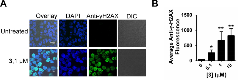
Figure 3:
Effect of 3 on DNA damage. A, representative confocal microscopy images of the effect of 3 on DNA damage in MDA-MB-231 cells by staining for γ-H2AX foci. White scale bar indicates 20 μm. B, quantification of DNA damage upon treatment of MDA-MB-231 cells with 3. * indicates p<0.5; ** indicates p<0.01, as determined by a two-tailed Student t test. See also Figure S4.

