. 2018 Oct 31;8(1):90–100. doi: 10.1039/c8tx00181b

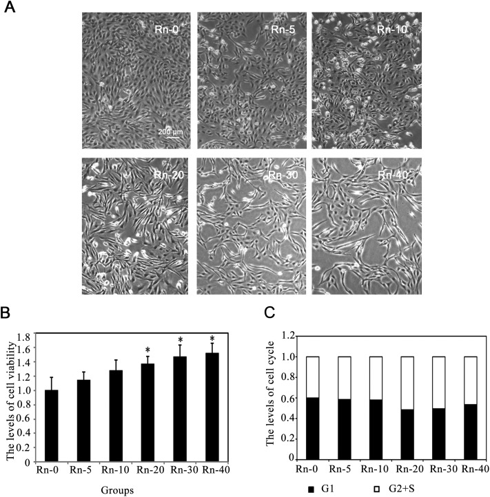
Fig. 1. The morphological changes of lung epithelial cells caused by radon exposure. (A) Followed byradon exposure for the indicated times, the morphology of BEAS-2B cells was captured. The cells were divided into control group (Rn-0) and radon exposure groups [(Rn-5) group of cells with 5 times radon exposure; (Rn-10) group of cells with 10 times radon exposure; (Rn-20)group of cells with 20 times radon exposure; (Rn-30) group of cells with 30 times radon exposure; (Rn-40) group of cells with 40 times radon exposure.] Representative cell images from each group are shown. (B) Radon exposure up-regulated the cell proliferation in a time dependent manner. BEAS-2B cells from each groups were harvested. The levels of cell viability were determined by MTT assay for 72 h. Results are expressed as means ± SD (n = 4). (*p < 0.05 radon exposure groups vs. control group). (C) Radon exposure did not affect the cell cycle. BEAS-2B cells from each groups were harvested. The levels of cell cycle were determined.

Fig. 1