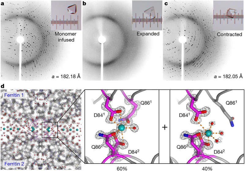
Fig. 3 |.
Atomic-level structural characterization of ferritin crystal-hydrogel hybrids by XRD. a-c, XRD patterns (at temperature T = 293 K) of a ferritin crystal infused with polymer precursors (a), after polymerization and expansion (b) and after contraction with CaCl2 (c). Light micrographs of the crystal are shown in the insets; the separation between the major ticks of the ruler is 100 μm. d, 1.06-Å-resolution structure (T = 100 K; PDB ID, 6B8F) of the contracted ferritin crystal-hydrogel hybrid, showing the electron density surrounding the Ca-mediated ferritin-ferritin interfaces and highlighting the two observed Ca coordination conformations. The electron density (2Fo-Fc) map (grey) is contoured at 1.5σ. Water molecules and Ca ions are shown as red and blue spheres, respectively.

