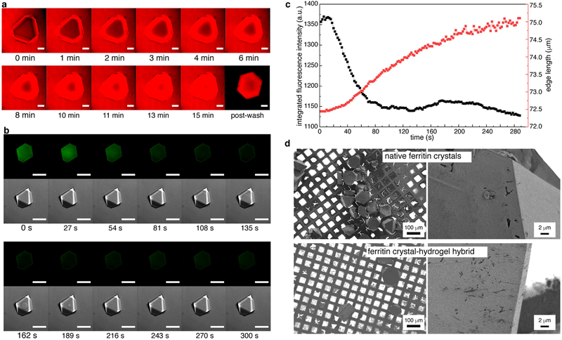
Extended Data Fig. 2 |. Molecular diffusion and polymerization in ferritin crystals, monitored using confocal microscopy.
a, Diffusion of rhodamine B into a ferritin crystal over 15 min. b, c, In crystallo polymerization of the hydrogel network, monitored through the decrease of integrated pyranine fluorescence (green fluorescence channel). The corresponding bright-field (DIC) images show the diffusion of the aqueous NaCl solution into the crystal. The ring-shaped diffusion front becomes evident at time t = 108 s and disappears by t = 216 s. The crystal expands by approximately 5% (edge length) during polymerization. Scale bars in a and b correspond to 100 μm. d, Scanning electron microscopy images of native ferritin crystals (top) and crystal-hydrogel hybrids (bottom).

