. 2018 Oct 9;10(2):398–405. doi: 10.1039/c8sc04266g

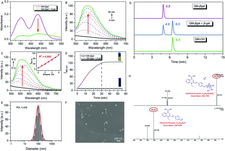
Fig. 2. (A) Absorption and (B) time-dependent fluorescence spectra of QM–βgal (10 μM) with 6 U β-gal in aqueous solution (PBS/DMSO = 95 : 5, v/v, 50 mM, pH = 7.4) at 37 °C. (C) Fluorescence spectra of QM–βgal (10 μM) upon treatment with increasing concentrations of β-gal (0–6 U). Inset (C): fluorescence intensity I560 nm as a function of β-gal concentration. (D) Fluorescence intensity I560 nm as a function of time, λex = 434 nm. (E) DLS data and (F) SEM of the ensemble system of QM–βgal and β-gal. (G) Reverse-phase HPLC chromatogram of QM–βgal, QM–βgal reaction with β-gal for 20 min, and QM–OH. The eluent is methanol/H2O (v/v, 8 : 2) mixed solvent. The flow rate is 0.6 mL min–1, and the detection wavelength is 434 nm. (H) HRMS demonstrating the enzyme-activatable mechanism and showing the cleavage product QM–OH.

Fig. 2